Rabu, 29 Juli 2009


Diabetes Mellitus, Type 2

Author: Jean-Claude DesMangles, MD, Assistant Professor, Department of Pediatrics, Creighton University School of Medicine
Contributor Information and Disclosures

Updated: Jul 2, 2009

Introduction

Background

Until recently, type 2 diabetes mellitus was almost exclusively a disease of adults. Coinciding with the increasing prevalence of obesity among American children, the incidence of type 2 diabetes in children and adolescents has markedly increased to the point that it accounts for as many as one third of all the new cases of diabetes diagnosed in adolescents. This trend is particularly pronounced in minority racial and ethnic groups. border=

Pathophysiology

In individuals without diabetes, approximately 50% of their total daily insulin is secreted during basal periods to suppress lipolysis, proteolysis, and glycogenolysis. In response to a meal, rapid insulin secretion (also called first-phase insulin secretion) ensues. This secretion facilitates the peripheral utilization of the prandial nutrient load, suppresses hepatic glucose production, and limits postprandial elevations in glucose levels. The second phase of insulin secretion follows and is sustained until normoglycemia is restored.

Type 2 diabetes spans a continuum from impaired glucose tolerance and impaired fasting glucose to frank diabetes resulting from progressive deterioration of both insulin secretion and action. Although the first phase of insulin response is markedly reduced early in the course of the disease, ongoing disorganized insulin secretion associated with deterioration of peripheral insulin action occurs during the progression from normal to impaired glucose tolerance to frank diabetes.

In parallel, as a result of decreased insulin sensitivity in the liver, endogenous glucose output increase adds to the already hyperglycemic milieu, worsening both peripheral insulin resistance and beta-cell function. Failure of the beta cell to keep up with the peripheral insulin resistance is the basis for the progression from impaired glucose tolerance to overt clinical type 2 diabetes. A longitudinal study demonstrated that, during the transition between normal glucose tolerance to diabetes, 31% of the person's insulin-mediated glucose disposal capacity is lost, whereas 78% of the acute insulin response is also lost during the same period.

Frequency

United States

Although type 2 diabetes is widely diagnosed in adults, its frequency has increased markedly in the pediatric age group during the past decade. Type 2 diabetes represents 8-45% of all new cases of diabetes reported among children and adolescents. Most pediatric patients in whom type 2 diabetes is diagnosed belong to minority communities.

International

An increased prevalence of type 2 diabetes has also been recognized in countries other than the United States, including Japan, where the incidence has doubled during the past 2 decades. In the Chinese, Taiwanese, and indigenous people of Australia, a trend for type 2 diabetes to occur at younger ages than before has also been recognized.

Mortality/Morbidity

Overall, morbidity and mortality associated with type 2 diabetes are related to short- and long-term complications.

  • According to a follow-up study of Pima Indians in whom type 2 diabetes was diagnosed before the age of 20 years, the incidence of nephropathy was not significantly different from that in patients with adult-onset diabetes. This result indicated a high risk of end-stage renal disease in the third and fourth decades of life.
  • In a comparative study among youths with type 1 and type 2 diabetes, the cumulative incidence of nephropathy was higher than it was in those with type 1 diabetes. Nephropathy also appeared earlier in type 2 than in type 1 diabetes.
  • The risk of retinopathy is lower in patients with youth-onset type 2 diabetes than in those with adult-onset diabetes.

Race

Type 2 diabetes primarily affects minority populations.

  • From 1967-1976 to 1987-1996, the prevalence of type 2 diabetes increased 6-fold in Pima Indian adolescents and appeared for the first time in children and adolescents younger than 15 years.
  • Similar increases in prevalence were observed among Japanese, Asian-American, and African-American children. In several clinics across the United States, pediatric patients with a diagnosis of type 2 diabetes were from minority ethnic groups (African-American, Asian, and Hispanic groups).

Sex

The prevalence of type 2 diabetes in the pediatric population is higher among girls than boys, just as it is higher among women than men.

Age

The mean age of onset of type 2 diabetes is 12-16 years; this period coincides with puberty, when a physiologic state of insulin resistance develops. In this physiologic state, type 2 diabetes develops only if inadequate beta-cell function is associated with other risk factors (eg, obesity).

Clinical

History

At the time of diagnosis, determine whether a patient has type 1 or type 2 diabetes because patients with type 1 diabetes are totally dependent on exogenous insulin administration for survival, whereas patients with type 2 diabetes do not necessarily require exogenous insulin to survive.

  • Because of the increasing prevalence of obesity in the pediatric population, the percentage of immune-mediated diabetes in overweight or obese patients is increasing, rendering the distinction between type 1 and type 2 diagnoses difficult at times. Blood glucose monitoring is required regardless of the type of diabetes, and treatment with insulin should be started when indicated.
  • The onset of type 2 diabetes is usually slow and insidious; it most often occurs in overweight or obese patients from a minority group.
    • Patients with type 2 diabetes often have signs of insulin resistance, such as hypertension or acanthosis nigricans.
    • A strong family history for the disease is usually reported among affected youth. The families of adolescents with type 2 diabetes also often have lifestyle risk factors leading to obesity.
    • Children with diabetes type 2 are more likely to report a family history of cardiovascular disease.
    • Autoimmune markers are usually negative.
  • Type 1 diabetes occurs in people of all races; its onset is typically acute and severe.
    • Patients with type 1 diabetes are often lean and do not show manifestations of insulin resistance.
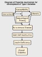
    • Autoimmunity is present in diabetes type 1.

Physical

  • Obesity is strongly associated with type 2 diabetes in children and adolescents. Eighty-five percent of children with type 2 diabetes are either overweight or obese (defined as at or above the 85th percentile of the sex-specific body mass index [BMI] for age-based growth charts).
  • Acanthosis nigricans, a marker of insulin resistance, is a velvety hyperpigmented thickening of the skin; it is frequently seen on the nape of the neck and in intertriginous areas; it is found in as many as 90% of children with type 2 diabetes.
  • Polycystic ovarian syndrome (PCOS) is a reproductive disorder commonly seen in young women with acanthosis nigricans. It is characterized by hyperandrogenism and chronic anovulation. The role of insulin resistance in the etiology of PCOS has been extensively studied, and medications that decrease insulin resistance and/or hyperinsulinemia in women with this syndrome often attenuate the hyperandrogenism and metabolic abnormalities.
  • Hypertension may occur in children with type 2 diabetes. The risk of macrovascular and microvascular diabetic complications is positively associated with elevated systolic blood pressure.
  • Ophthalmologic examination should be performed at or shortly after diagnosis to detect incipient retinopathy.

Causes

The major risk factors for type 2 diabetes in youths are the following:

  • Obesity and inactivity, which are important contributors to insulin resistance
  • Native American, Hispanic, Asian, and Pacific Islander descent
  • Family history of type 2 diabetes
  • Age of 12-16 years, the mean age at the onset of type 2 diabetes in youths (This age coincides with relative insulin resistance that occurs during pubertal development.)
  • Low birth weight

Diabetes Mellitus, Type 1

Author: William H Lamb, MBBS, MD, FRCP(Edin), FRCP, Clinical Lecturer, Department of Child Health, The General Hospital, Bishop Auckland, UK
Contributor Information and Disclosures

Updated: Jul 2, 2009

Introduction

Background

Diabetes mellitus (DM) is a chronic metabolic disorder caused by an absolute or relative deficiency of insulin, an anabolic hormone. Insulin is produced by the beta cells of the islets of Langerhans located in the pancreas, and the absence, destruction, or other loss of these cells results in type 1 diabetes (insulin-dependent diabetes mellitus [IDDM]). Most children with diabetes have type 1 diabetes mellitus (T1DM) and a lifetime dependence on exogenous insulin.

Possible mechanism for development of type 1 diab...

Possible mechanism for development of type 1 diabetes.

Possible mechanism for development of type 1 diab...

Possible mechanism for development of type 1 diabetes.

Type 2 diabetes mellitus (non–insulin-dependent diabetes mellitus [NIDDM]) is a heterogeneous disorder. Most patients with type 2 diabetes mellitus have insulin resistance, and their beta cells lack the ability to overcome this resistance. Although this form of diabetes was previously uncommon in children, in some countries, 20% or more of new patients with diabetes in childhood and adolescence have type 2 diabetes mellitus, a change associated with increased rates of obesity. Other patients may have inherited disorders of insulin release, leading to maturity onset diabetes of the young (MODY) or congenital diabetes.1,2

This topic addresses only type 1 diabetes mellitus.

Pathophysiology

Insulin is essential to process carbohydrates, fat, and protein. Insulin reduces blood glucose levels by allowing glucose to enter muscle cells and by stimulating the conversion of glucose to glycogen (glycogenesis) as a carbohydrate store. Insulin also inhibits the release of stored glucose from liver glycogen (glycogenolysis) and slows the breakdown of fat to triglycerides, free fatty acids, and ketones. It also stimulates fat storage. Additionally, insulin inhibits the breakdown of protein and fat for glucose production (gluconeogenesis) in both liver and kidneys.

Hyperglycemia (ie, random blood glucose concentration more than 200 mg/dL or 11 mmol/L) results when insulin deficiency leads to uninhibited gluconeogenesis and prevents the use and storage of circulating glucose. The kidneys cannot reabsorb the excess glucose load, causing glycosuria, osmotic diuresis, thirst, and dehydration. Increased fat and protein breakdown leads to ketone production and weight loss. Without insulin, a child with type 1 diabetes mellitus wastes away and eventually dies due to diabetic ketoacidosis (DKA).

An excess of insulin prevents the release of glucose into the circulation and results in hypoglycemia (blood glucose concentrations of <60>

Frequency

United States

The overall annual incidence has risen from approximately 16 cases per 100,000 population in the 1990s to 24.3 per 100,000 population currently and is probably still increasing. Although most new diabetes cases are type 1 (approximately 15,000 annually), increasing numbers of older children are being diagnosed with type 2 diabetes mellitus, especially among minority groups (3700 annually).3

International

Type 1 diabetes mellitus has wide geographic variation in incidence and prevalence.4 Annual incidence varies from 0.61 cases per 100,000 population in China, to 41.4 cases per 100,000 population in Finland. Substantial variations are observed between nearby countries with differing lifestyles, such as Estonia and Finland, and between genetically similar populations, such as those in Iceland and Norway. Even more striking are the differences in incidence between mainland Italy (8.4 cases per 100,000 population) and the Island of Sardinia (36.9 cases per 100,000 population). These variations strongly support the importance of environmental factors in the development of type 1 diabetes mellitus. Most countries report that incidence rates have at least doubled or more in the last 20 years. Incidence appears to increase with distance from the equator.5

Mortality/Morbidity

Information on mortality rates is difficult to ascertain without complete national registers of childhood diabetes, although age-specific mortality is probably double that of the general population.6,7 Children aged 1-4 years are particularly at risk and may die due to DKA at the time of diagnosis. Adolescents are also a high-risk group. Most deaths result from delayed diagnosis or neglected treatment and subsequent cerebral edema during treatment for DKA, although untreated hypoglycemia also causes some deaths. Unexplained death during sleep may also occur.8

The complications of type 1 diabetes mellitus can be divided into 3 major categories: acute complications, long-term complications, and complications caused by associated autoimmune diseases.

  • Acute complications reflect the difficulties of maintaining a balance between insulin therapy, dietary intake, and exercise. Acute complications include hypoglycemia, hyperglycemia, and DKA.
  • Long-term complications arise from the damaging effects of prolonged hyperglycemia and other metabolic consequences of insulin deficiency on various tissues. Although long-term complications are rare in childhood, maintaining good control of diabetes is important to prevent complications from developing in later life.9 The likelihood of developing complications appears to depend on the interaction of factors such as metabolic control, genetic susceptibility, lifestyle (eg, smoking, diet, exercise), pubertal status, and gender.10 Long-term complications include the following:
    • Retinopathy
    • Cataracts
    • Hypertension
    • Progressive renal failure
    • Early coronary artery disease
    • Peripheral vascular disease
    • Neuropathy, both peripheral and autonomic
    • Increased risk of infection
  • Associated autoimmune diseases are common with type 1 diabetes mellitus, particularly in children who have the human leukocyte antigen DR3 (HLA-DR3). Some conditions may precede development of diabetes; others may develop later. As many as 20% of children with diabetes have thyroid autoantibodies.

Race

Different environmental effects on type 1 diabetes mellitus development complicate the influence of race, but racial differences are evident. Whites have the highest reported incidence, whereas Chinese individuals have the lowest. Type 1 diabetes mellitus is 1.5 times more likely to develop in American whites than in American blacks or Hispanics. Current evidence suggests that when immigrants from an area with low incidence move to an area with higher incidence, their rates of type 1 diabetes mellitus tend to increase toward the higher level.

Sex

The influence of sex varies with the overall incidence rates. Males are at greater risk in regions of high incidence, particularly older males, whose incidence rates often show seasonal variation. Females appear to be at a greater risk in low-incidence regions.

Age

Generally, incidence rates increase with age until mid-puberty then decline after puberty, but type 1 diabetes mellitus can occur at any age. Onset in the first year of life, although unusual, can occur and must be considered in any infant or toddler because these children have the greatest risk for mortality if diagnosis is delayed. Their symptoms may include the following:

  • Severe monilial diaper/napkin rash
  • Unexplained malaise
  • Poor weight gain or weight loss
  • Increased thirst
  • Vomiting and dehydration, with a constantly wet napkin/diaper

Neonatal diabetes, including diagnosis in infants younger than 6 months, is most likely due to an inherited defect of the iKir6.2 subunit potassium channel of the islet beta cells, and genetic screening is indicated.11

In areas with high prevalence rates, a bimodal variation of incidence has been reported that shows a definite peak in early childhood (ie, 4-6 y) and a second, much greater peak of incidence during early puberty (ie, 10-14 y).12

Clinical

History

The most easily recognized symptoms of type 1 diabetes mellitus (T1DM) are secondary to hyperglycemia, glycosuria, and ketoacidosis (KA).

  • Hyperglycemia: Hyperglycemia alone may not cause obvious symptoms, although some children report general malaise, headache, and weakness. They may also appear irritable and become ill-tempered. The main symptoms of hyperglycemia are secondary to osmotic diuresis and glycosuria.
  • Glycosuria: This condition leads to increased urinary frequency and volume (eg, polyuria), which is particularly troublesome at night (eg, nocturia) and often leads to enuresis in a previously continent child. These symptoms are easy to overlook in infants because of their naturally high fluid intake and diaper/napkin use.
  • Polydipsia: Increased thirst, which may be insatiable, is secondary to the osmotic diuresis causing dehydration.
  • Weight loss: Insulin deficiency leads to uninhibited gluconeogenesis, causing breakdown of protein and fat. Weight loss may be dramatic, although the child's appetite usually remains good. Failure to thrive and wasting may be the first symptoms noted in an infant or toddler and may precede frank hyperglycemia.
  • Nonspecific malaise: Although this condition may be present before symptoms of hyperglycemia, or as a separate symptom of hyperglycemia, it is often only retrospectively recognized.
  • Symptoms of ketoacidosis
    • Severe dehydration
    • Smell of ketones
    • Acidotic breathing (ie, Kussmaul respiration), masquerading as respiratory distress
    • Abdominal pain
    • Vomiting
    • Drowsiness and coma
  • Other nonspecific findings
    • Hyperglycemia impairs immunity and renders a child more susceptible to recurrent infection, particularly of the urinary tract, skin, and respiratory tract.
    • Candidiasis may develop, especially in groin and flexural areas.

Physical

  • Apart from wasting and mild dehydration, children with early diabetes have no specific clinical findings.
  • A physical examination may reveal findings associated with other autoimmune endocrinopathies, which have a higher incidence in children with type 1 diabetes mellitus (eg, thyroid disease with symptoms of overactivity or underactivity and possibly a palpable goiter).
  • Cataracts are rarely presenting problems and typically occur in girls with a long prodrome of mild hyperglycemia.
  • Necrobiosis lipoidica usually, but not exclusively, occurs in people with diabetes. Necrobiosis most often develops on the front of the lower leg as a well-demarcated, red, atrophic area. The condition is associated with injury to dermal collagen, granulomatous inflammation, and ulceration. The cause of necrobiosis is unknown, and the condition is difficult to manage. It is also associated with poor metabolic control and a greater risk of developing other diabetes-related complications.

Causes

Most cases (95%) of type 1 diabetes mellitus are the result of environmental factors interacting with a genetically susceptible person. This interaction leads to the development of autoimmune disease directed at the insulin-producing cells of the pancreatic islets of Langerhans. These cells are progressively destroyed, with insulin deficiency usually developing after the destruction of 90% of islet cells.

  • Genetic issues
    • Clear evidence suggests a genetic component in type 1 diabetes mellitus.
    • Monozygotic twins have a 60% lifetime concordance for developing type 1 diabetes mellitus, although only 30% do so within 10 years after the first twin is diagnosed. In contrast, dizygotic twins have only an 8% risk of concordance, which is similar to the risk among other siblings.
    • The frequency of diabetes developing in children with a diabetic mother is 2-3% and 5-6% if the father has type 1 diabetes mellitus. The risk to children rises to almost 30% if both parents are diabetic.
    • Human leukocyte antigen (HLA) class II molecules DR3 and DR4 are associated strongly with type 1 diabetes mellitus. More than 90% of whites with type 1 diabetes mellitus express one or both of these molecules, compared with 50-60% in the general population.
    • Patients expressing DR3 are also at risk for developing other autoimmune endocrinopathies and celiac disease. These patients are more likely to develop diabetes at a later age, to have positive islet cell antibodies, and to appear to have a longer period of residual islet cell function.
    • Patients expressing DR4 are usually younger at diagnosis and more likely to have positive insulin antibodies, yet they are unlikely to have other autoimmune endocrinopathies.
    • The expression of both DR3 and DR4 carries the greatest risk of type 1 diabetes mellitus; these patients have characteristics of both the DR3 and DR4 groups.
  • Environmental factors
    • Environmental factors are important because even identical twins have only a 30-60% concordance for type 1 diabetes mellitus and because incidence rates vary in genetically similar populations under different living conditions.13
    • No single factor has been identified, but infections and diet are considered the 2 most likely environmental candidates.
    • Viral infections may be the most important environmental factor in the development of type 1 diabetes mellitus,14 probably by initiating or modifying an autoimmune process. Instances have been reported of a direct toxic effect of infection in congenital rubella. One survey suggests enteroviral infection during pregnancy carries an increased risk of type 1 diabetes mellitus in the offspring. Paradoxically, type 1 diabetes mellitus incidence is higher in areas where the overall burden of infectious disease is lower.
    • Dietary factors are also relevant. Breastfed infants have a lower risk for insulin-dependent diabetes mellitus (IDDM), and a direct relationship is observed between per capita cow's milk consumption and the incidence of diabetes. Some cow's milk proteins (eg, bovine serum albumin) have antigenic similarities to an islet cell antigen. Nitrosamines, chemicals found in smoked foods and some water supplies, are known to cause type 1 diabetes mellitus in animal models; however, no definite link has been made with humans.
    • The known association of increasing incidence of type 1 diabetes mellitus with distance from the equator may now have an explanation. Reduced exposure to UV light and lower vitamin D levels, both of which are more likely found in the higher latitudes, are associated with an increased risk of type 1 diabetes mellitus.15
  • Chemical causes: Streptozotocin and RH-787, a rat poison, selectively damage islet cells and can cause type 1 diabetes mellitus.
  • Other causes

diabetes insipidus


Author: James CM Chan, MD, Professor of Pediatrics, University of Vermont College of Medicine; Director of Research, The Barbara Bush Children's Hospital, Maine Medical Center
Coauthor(s): Karl S Roth, MD, Professor and Chair, Department of Pediatrics, Creighton University School of Medicine

Updated: Feb 6, 2009

Introduction

Background

The word diabetes is derived from the Greek verb diabainein, which means to stand with legs apart (as in urination) or to go through. Insipidus comes from a Latin word meaning without taste. In contrast to diabetes mellitus (DM), which describes the excretion of sweet urine, diabetes insipidus (DI) describes the passing of tasteless urine because of its relatively low sodium content.

Nephrogenic diabetes insipidus (NDI) reached North America in 1761 and was carried by Ulster Scots who arrived in Nova Scotia, Canada, on a ship named Hopewell.1 Scottish folklore reports the existence of the disease in Scotland before 1761. According to legend, a gypsy woman traveling with her thirsty son is denied water by a housewife. The gypsy woman curses the housewife, causing the housewife's sons to crave water while condemning her daughters to pass the curse on to future generations.

Pathophysiology

The basis of water loss in diabetes insipidus is distinct from water loss caused by diabetes mellitus. The renal tubular collecting ducts are unable to concentrate urine secondary to vasopressin deficiency or resistance. The collecting duct concentrates urine by reabsorbing water, a function controlled by the posterior pituitary gland via secretion of vasopressin or antidiuretic hormone (ADH). Reabsorption of sugars, amino acids, and virtually all electrolytes is completed by the time the urine has reached this segment of the nephron. Consequently, the inability to conserve water by reabsorption in the collecting duct depletes body water but leaves sodium unaffected. The net result is an extremely diluted, increased urine output resulting in hypernatremia. Polydipsia follows, as the thirst mechanism urges replenishment of body water.

Secretion of vasopressin occurs in the posterior pituitary gland and is regulated at the paraventricular and supraoptic nuclei, which sense changes in osmolality. Destruction of the paraventricular or supraoptic nuclei or of the posterior pituitary by tumor, pressure, or surgical ablation results in decreased vasopressin secretion and central diabetes insipidus (CDI). Alternatively, diabetes insipidus may be idiopathic or inherited either as an autosomal dominant or as an autosomal recessive trait (locus 20p13).

Nephrogenic diabetes insipidus arises from defective or absent receptor sites at the cortical collecting duct segment of the nephron (X-linked, vasopressin V2 receptor deficiency, locus Xq28) or defective or absent aquaporin, the protein that transports water at the collecting duct (autosomal recessive, locus 12q13). The X-linked variety of nephrogenic diabetes insipidus (NDI) accounts for about 90% of all such cases.

As a consequence of one of these defects, the ducts do not appropriately respond to vasopressin. Normally, vasopressin is transported in the blood to receptor sites on the basolateral surface of the collecting duct membrane. Through a G protein–adenylate cyclase coupling, activation of the vasopressin receptor increases cyclic adenosine monophosphate (AMP) production and stimulates protein kinase A, leading to increased recycling of the protein aquaporin in the plasma membrane.

In the presence of vasopressin stimulus, exocytic insertion of aquaporin into the apical, or luminal, surface of the tubule cell occurs. Aquaporin enhances water entry into the cell from the lumen. Absence of the vasopressin receptor does not allow this process to take place, causing inhibition of water uptake and polyuria. Alternatively, defective or absent aquaporin impairs the process in the presence of normal V2 receptors.

Frequency

United States

Tumors, infiltrative lesions, malformations, and neurosurgical procedures are the most common causes of central diabetes insipidus. Of the genetic etiologies, the overall incidence in the general population is estimated to be 3 cases per 100,000 population (0.003%). The male-to-female ratio is 60:40. X-linked nephrogenic diabetes insipidus is very rare, although it exceeds the recessive variety by a ratio of 9:1. The mutation for males is 4 cases per million population.

Mortality/Morbidity

Dehydration results from an inability to reabsorb free water at a site distal to electrolyte reabsorption. Any patient unable to continuously replace water loss is vulnerable to dehydration, especially in warm weather when insensible water loss through perspiration and respiration substantially increases risk. Electrolyte abnormalities are caused by the loss of urinary free water, which produces hyperosmolar dehydration, leading to hypernatremia, hyperchloremia, and prerenal azotemia. Diminished blood volume increases blood viscosity and the risk of sludging and thrombosis.

Failure to thrive occurs because of the patient's constant thirst conferring a sense of fullness that offsets the sense of hunger. The affected individual eats less than necessary for normal growth. Seizures are a consequence of the electrolyte abnormalities introduced in the CNS by severe hypernatremia and hyperosmolar dehydration. Mental retardation results from the damage to the CNS caused by severe hyperosmolarity, seizures, and potential hypoxia, all of which are thought to account for the frequent occurrence of mental retardation. Death can occur from a hypovolemic shock or a hypernatremic seizure.

Sex

Central diabetes insipidus secondary to hypothalamic-pituitary lesions occurs at random and should, therefore, be evenly distributed between the sexes. Autosomal dominant and autosomal recessive central diabetes insipidus occur equally in both sexes. Nephrogenic diabetes insipidus caused by an X-linked mutation affects only males. Autosomal dominant and autosomal recessive forms of nephrogenic diabetes insipidus equally affect both sexes.

Age

Diabetes insipidus occurs in people of a wide age range. Children who present with autosomal recessive central diabetes insipidus are generally younger than 1 year. Children who present with autosomal dominant central diabetes insipidus are often older than 1 year. Nephrogenic diabetes insipidus forms (including X-linked, autosomal dominant, and autosomal recessive forms) develop in early infancy, often in neonates younger than 1 week.

Clinical

History

  • Diagnosis of diabetes insipidus (DI) may be difficult in infants and children because of nonspecific presenting features (eg, poor feeding, failure to thrive, irritability). Therefore, a high index of suspicion is necessary.
  • The earliest signs of diabetes insipidus include a vigorous suck with vomiting, fever without apparent cause, constipation, and excessively wet diapers from urination.
  • In older infants and young children, irritability is generally due to a borderline state of dehydration coupled with hypernatremia and, sometimes, fever.
  • Nocturia is common and expected because of increased urine production.
  • Central diabetes insipidus (CDI) tends to suddenly develop.

Physical

  • The typical examination reveals an irritable infant with a dripping wet diaper, along with detectable signs of dehydration (eg, dry mucous membranes, diminished skin turgor, decreased tearing, tachycardia). Often, skin turgor is not diminished in individuals with hypernatremic dehydration despite significant dehydration.
  • In severely dehydrated patients, the pulse may be thready and rapid. Hypotension may be present because of hypovolemic shock.
  • Mobile fecaliths may be palpable in the abdomen.

Causes

Diabetes insipidus is due to either (1) deficiency of vasopressin secretion by the pituitary gland (central diabetes insipidus or neurogenic diabetes insipidus) or to (2) renal tubular unresponsiveness to vasopressin (nephrogenic diabetes insipidus [NDI]).

  • Nongenetic causes
    • Typical injuries include head trauma, tumor, and neurosurgical procedures.
    • At all ages, destructive lesions of the pituitary and/or hypothalamus are the most common cause of diabetes insipidus.
  • Genetic causes
    • Central diabetes insipidus with an autosomal dominant pattern inheritance is due to a mutation in the prepro-arginine vasopressin (prepro-AVP2) gene, mapped to locus 20p13.
    • Central diabetes insipidus with diabetes mellitus, optic atrophy, and mental retardation (Wolfram syndrome) may be inherited in an autosomal recessive pattern (locus 4p16) or may be due to mitochondrial deletions.
    • X-linked nephrogenic diabetes insipidus occurs from mutations in the antidiuretic arginine vasopressin V2 receptor (AVPR2) gene, mapped to Xq28.2,3,4
    • Nephrogenic diabetes insipidus with an autosomal dominant or recessive pattern is due to mutations in the gene designated AQP2; this gene directs water channel formation in the distal membrane and has not yet been mapped.


laporan kimia


LAPORAN PRAKTIKUM KIMIA

Nama :

Kelas :

No :

Judul : Sel volta

Tanggal :

Tujuan :-Dapat merangkaikan sel volta

-Dapat menentukan Esel secara teori

-Dapat menentukan Esel secara eksperimen

Alat & Bahan :-Gelas kimia 100 mL

-Voltmeter

-Kabel / jepit buaya

-Elektroda seng,tembaga,magnesium,besi

-Jembatan garam

-Larutan CuSO4

-Larutan ZnSO4

-Larutan MgSO4

-Larutan FeSO4

Urutan Kerja :

  1. Pasang alat
  2. Isilah gelas kimia dengan larutan CuSO4 & ZnSO4 1 molar
  3. Celupkan sepotong seng dalam ZnSO4
  4. Celupkan sepotong tembaga dalam CuSO4
  5. Hubungkan kedua larutan dengan jembatan garam
  6. Hubungan kedua lempeng logam melalui voltmeter.jika jarum bergerak kea rah negatife, segera putuskan dan jika jarum voltmeter kearah positif biarkan dan baca beda potensialnya,catat beda potensialnya pada table berikut ini:
  7. No

    + setengah sel katode

    A

    B

    C

    D


    - setengah sel anode

    Cu2+/ Cu

    Zn2+ / Zn

    Mg2+ / Mg

    Fe2+ / Fe

    1

    2

    3

    4

    Cu / Cu2+

    Zn / Zn2+

    Mg / Mg2+

    Fe / Fe2+

    ----------

    + 0,85

    + 0,8

    + 0.65

    - 0,85

    ---------------

    - 0,625

    ---------------

    -0,8

    + 0,625

    ---------------

    - 0,31

    -0,65

    ---------------

    + 0,31

    ---------------

    Jawaban pertanyaan:

    1. Tulislah bagan sel serta persamaan reaksi setengah sel dan reaksi untuk soal

    3 –A,3-B,dan 2-A dalam tabel eksperimen:

    # 3 –A : Mg / Mg2+ // Cu2+ / Cu

    Anoda : Mg Mg2+ + 2e Eo = -2,37 V

    Katoda:Cu2++ 2e Cu Eo = +0,34 V


    Reaksi sel: Cu2+ + Mg Cu + Mg2+ Esel = +2,71 V

    # 3-B : Mg / Mg2+ // Zn2+ / Zn

    Anoda : Mg Mg2+ + 2e Eo = -2,37 V

    Katoda: Zn2+ + 2e Zn Eo = -0,76 V


    Reaksi sel: Zn2+ + Mg Zn + Mg2+ Esel= 71,61V

    # 2-A : Zn / Zn2+// Cu2+ / Cu

    Anoda : Zn Zn2+ + 2e Eo = -0.76 V

    Katoda: Cu2+ + 2e Cu Eo = +0.34 V

    Reaksi sel: Cu2+ + Zn Cu + Zn2+ Esel=+1,1 V

    2) Hitunglah potensial sel 2-A berdasarkan petensial sel 3-A dan potensial sel 3-Byang tercantum dalam table experiment !

    Mg / Mg2+ // Cu2+ / Cu Eo = + 0.8 V

    Mg / Mg2+ // Zn2+ / Zn Eo = -0.625 V

    Jawab Mg / Mg2+ // Cu2+ / Cu Eo = + 0.8 V

    Mg / Mg2+ // Zn2+ / Zn Eo = + 0.625 V

    Zn / Zn2+// Cu2+ / Cu Eo = + 1,425 V


PIETER'S WEBLOG Blak Magik is Designed by productive dreams for smashing magazine Bloggerized by Ipiet Blogger Templates © 2008